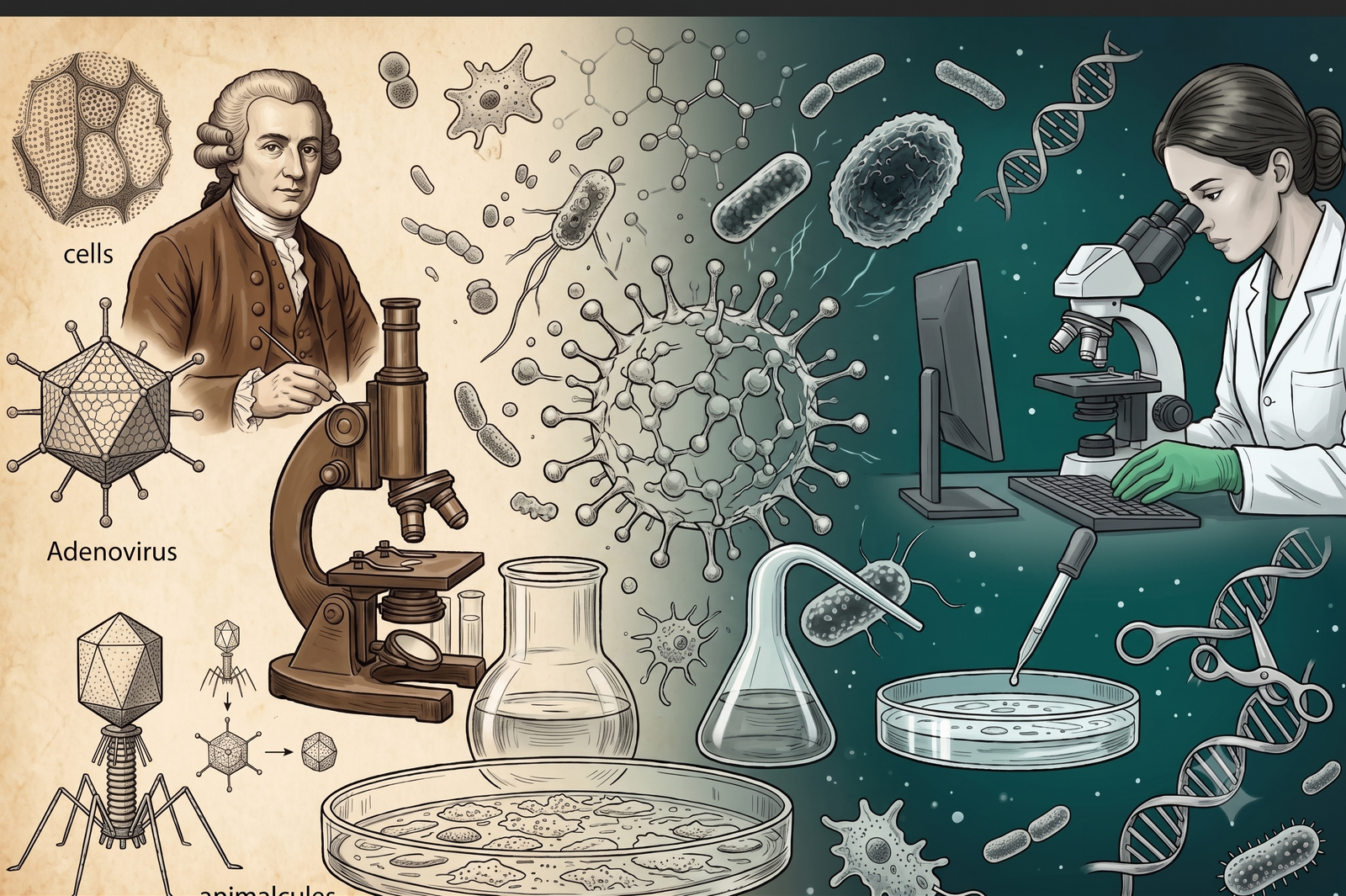
The history of biology is fundamentally divided into two eras: the era of the visible and the era of the invisible. For millennia, humanity’s understanding of life, disease, and decay was bound to what the naked eye could perceive. The advent of microbiology—the study of organisms too small to be seen without magnification—shattered these sensory boundaries.
It revealed a bustling, complex microscopic universe that dictates the survival, evolution, and death of all macroscopic life.
From the crude lens-grinding of the seventeenth century to the precise gene-editing technologies of the present era, the advances in microbiology have fundamentally rewritten the rules of medicine, agriculture, industry, and biotechnology.
The Advent: Piercing the Veil of the Invisible
The birth of microbiology as a formal scientific endeavor can be traced back to two distinct seventeenth-century pioneers who pierced the veil of the hidden world:
Robert Hooke and Antonie van Leeuwenhoek
In 1665, the English polymath Robert Hooke published Micrographia, a groundbreaking work detailing his observations using a compound microscope. Hooke was the first to coin the term “cell” after examining the structural, box-like compartments of cork tissue.
A decade later, a Dutch draper named Antonie van Leeuwenhoek revolutionized lens-making. Using highly refined, single-lens microscopes of his own design, Leeuwenhoek observed scrapings from his teeth, pond water, and blood droplets. He became the first human to witness living, moving single-celled organisms—including bacteria, protozoa, and spermatozoa—which he poetically termed “animalcules” (little animals).
Overturning Spontaneous Generation
Despite Leeuwenhoek’s astonishing discovery, the scientific community did not immediately connect these tiny entities to disease or ecological cycles. For nearly two centuries, the dominant paradigm remained the theory of spontaneous generation—the belief that living organisms could arise spontaneously from non-living, decaying matter (such as maggots emerging spontaneously from rotting meat).
The Golden Age: Establishing Foundations and the Germ Theory
The true institutionalization of microbiology occurred during the “Golden Age of Microbiology” (roughly 1857 to 1914), dominated by two towering scientific figures whose fierce, parallel work laid the bedrock of modern medicine.
[ Pre-1860s ] ─────────────────► [ 1860s – 1880s ] ──────────────► [ 1928 – 1940s ]
• Spontaneous Generation • Pasteurization
• Fleming’s Penicillin
• Miasma Theory (“Bad Air”) • Germ Theory
Established • Industrial Scale Antibiotics
• Hooke/Leeuwenhoek Observ. • Koch’s Postulates
Implemented • Mass Drop in Infection Mortality
Louis Pasteur
The brilliant French chemist Louis Pasteur dealt the final death blow to spontaneous generation through his elegant swan-neck flask experiments. He proved that microbes do not spontaneously manifest; they are carried in the air and contaminate sterile environments. Pasteur’s subsequent discoveries transformed global industry and medicine.
He demonstrated that microscopic yeast cells drive fermentation, and developed pasteurization—a controlled heating process to destroy spoilage microbes in milk, beer, and wine.
Crucially, Pasteur went on to develop the foundational principles of immunization, creating the world’s first laboratory-developed vaccines for anthrax and rabies by using weakened (attenuated) strains of pathogens.
Robert Koch
While Pasteur was a visionary of immunization, the German physician Robert Koch was the master of isolation and definitive proof. Koch formulated a rigorous set of scientific criteria known as Koch’s Postulates, which provided a systematic framework to prove that a specific microbe causes a specific disease.
Using newly invented solid agar growth media, Koch successfully isolated the causative agents of anthrax (Bacillus anthracis), cholera (Vibrio cholerae), and tuberculosis (Mycobacterium tuberculosis). His work definitively established the Germ Theory of Disease, forever replacing ancient, superstitious beliefs like the “miasma theory” (the idea that diseases were caused by foul, noxious air).
The Twentieth Century: The Antibiotic Revolution and Molecular Biology
As microbiology entered the twentieth century, the focus shifted from identifying pathogens to conquering them and uncovering their internal genetic machinery.
The Magic Bullet and Penicillin
The pursuit of a chemical “magic bullet” that could kill harmful microbes inside a living host without damaging human tissue led to the birth of antimicrobial chemotherapy. In 1928, Scottish bacteriologist Alexander Fleming noticed that a stray mold contaminant (Penicillium notatum) had inhibited the growth of staphylococcus bacteria on a petri dish.
The subsequent isolation, purification, and mass industrial production of penicillin by Howard Florey and Ernst Chain during World War II marked the dawn of the antibiotic era. Diseases that had acted as absolute death sentences for generations—such as pneumonia, syphilis, and wound infections—became entirely curable overnight.
Microbes as Genetic Blueprints
Concurrently, microbiologists realized that bacteria and viruses, due to their simple structures and incredibly rapid reproduction cycles, were the ideal model organisms for studying the fundamental molecular mechanisms of life. In 1944, Oswald Avery, Colin MacLeod, and Maclyn McCarty used the bacterium Streptococcus pneumoniae to prove that DNA, not protein, is the hereditary material of cells.
Similarly, the study of bacterial viruses (bacteriophages) by researchers like Max Delbrück laid the foundation for modern molecular biology, unlocking the secrets of DNA replication, transcription, and genetic translation.
Contemporary Advances: Genomics, the Microbiome, and CRISPR
Today, microbiology has entered a hyper-advanced, technological era where scientists no longer just observe or destroy microbes; they sequence, engineer, and collaborate with them.
The Genomic Era and Next-Generation Sequencing
For over a century, microbiologists were limited by a frustrating reality: less than 1% of environmental microbes can be successfully grown (cultured) in a traditional laboratory setting. The advent of Metagenomics and Next-Generation Sequencing (NGS) completely bypassed this bottleneck.
By extracting and sequencing DNA directly from environmental or biological samples—such as soil, deep-sea vents, or the human gut—scientists can catalog entirely new domains of microbial life without ever growing a single cell.
The Human Microbiome
This genomic revolution has profoundly reshaped human physiology through the study of the Human Microbiome.
We now understand that the human body is a complex ecosystem populated by trillions of microbial cells that outnumber or match our own human cells. Research into the gut microbiome has revealed that these commensal bacteria are not passive passengers; they actively regulate our immune systems, synthesize critical vitamins, aid digestion, and directly influence our mental health and neurological functions via the gut-brain axis.
CRISPR-Cas9: Microscopic Defenses to Global Tools
Perhaps the most staggering modern advance derived from microbiology is CRISPR-Cas9 gene editing. Originally discovered as a primitive, adaptive immune defense system used by bacteria to fight off viral attacks, scientists re-engineered CRISPR into a highly precise molecular scissor.
This microbial defense mechanism has revolutionized modern medicine and biotechnology, allowing scientists to edit the genomes of plants, animals, and humans with unprecedented accuracy—opening the door to curing genetic diseases and engineering resilient crops.
Virology is the highly specialized branch of microbiology that deals with the study of viruses—their structure, classification, genetics, physiology, pathogenesis, and interaction with host organisms.
Unlike living bacteria, viruses occupy a fascinating biological gray zone: they are completely inert outside of a host cell, lacking the cellular machinery necessary to generate energy, synthesize proteins, or reproduce independently. Because of this, they are classified as obligate intracellular parasites.
1. Structural Anatomy of a Virus
At their most fundamental level, viruses are remarkably simple, elegant structures designed for a single purpose: to transport a genetic payload into a target cell.
Virology is a specialized branch of microbiology dedicated specifically to studying viruses, which are unique, non-cellular infectious agents that depend entirely on host cells to reproduce. Looking at the image, you can see four major virus structural types classified by their physical shapes.
Starting on the left, the helical type, shown by the tobacco mosaic virus, features an elongated capsid made of tiny subunits enclosing its genetic material. Moving to the second type, the icosahedral or polyhedral structure seen in the adenovirus displays a geometric, multi-sided shell equipped with projecting fibers.
The third structure is the enveloped virus, exemplified by the influenza virus, where the inner protein shell is wrapped inside an outer membrane envelope studded with special glycoproteins. Finally, on the far right, the complex type is represented by a bacteriophage, which targets bacteria using a distinct architecture consisting of a genetic head, a tail sheath, and leg-like tail fibers.
Unlocking these diverse viral forms is crucial for designing modern vaccines and cutting-edge gene therapies to combat global health crises.
Every standard viral particle (known as a virion) contains at least two primary components:
• Genetic Material (Core): Unlike cellular organisms which always use double-stranded DNA, viruses can use single-stranded DNA (ssDNA), double-stranded DNA (dsDNA), single-stranded RNA (ssRNA), or double-stranded RNA (dsRNA).
• Capsid: A protective protein shell that encloses the genetic core. The capsid is composed of repeating protein subunits called capsomeres.
Structural Types
• Helical: Rod-shaped or cylindrical capsids where the proteins wrap around the genetic material in a spiral (e.g., Tobacco Mosaic Virus).
• Icosahedral: A geometric, spherical-looking shape featuring 20 equilateral triangular faces (e.g., Adenovirus).
• Enveloped: Some viruses steal a piece of the host cell’s outer membrane as they exit. This creates a lipid envelope layer spiked with viral glycoproteins that help the virus trick and bind to the next host cell (e.g., Influenza, HIV, and SARS-CoV-2). Viruses without this layer are called naked viruses.
• Complex (Bacteriophage): Specialized viruses that hunt bacteria. They look like microscopic lunar landers, possessing an icosahedral head containing DNA, a hollow sheath tube, and leg-like tail fibers used to latch onto bacterial cell walls.
2. The Viral Replication Cycle
To replicate, a virus must hijack a host cell and force it to act as an assembly factory. This process follows six fundamental steps:
[ 1. Attachment ] ──► [ 2. Penetration ] ──► [ 3. Uncoating ]
│
[ 6. Release ] ◄── [ 5. Assembly ] ◄── [ 4. Biosynthesis ]
1. Attachment (Adsorption): The virus utilizes its surface proteins or spike glycoproteins to lock onto specific receptors on the surface of the host cell. This dictates its tropism (which specific cell types it can infect).
2. Penetration: The virus enters the cell, either by fusing its envelope with the host membrane or by triggering receptor-mediated endocytosis (where the host cell engulfs it).
3. Uncoating: The protective capsid shell dissolves, releasing the viral genome directly into the host cell’s cytoplasm or nucleus.
4. Biosynthesis: The viral genome hijacks the host’s enzymes and ribosomes. The cell stops making its own proteins and begins rapidly copying the viral genetic code and synthesizing new capsid proteins.
5. Assembly (Maturation): The freshly manufactured viral genomes and capsid proteins spontaneously snap together inside the cell to form thousands of new, complete virions.
6. Release: The new viruses exit the cell. Naked viruses typically cause lysis (bursting and destroying the host cell entirely). Enveloped viruses slip through the membrane via budding, taking pieces of the host membrane with them over time.
3. Why Viruses are Critical to Science and Medicine
The Threat of Pandemics and Emerging Viruses
Because many RNA viruses have incredibly high mutation rates and replicate rapidly, they can quickly bypass host immunity and adapt to new species (known as a zoonotic spillover). Understanding how these viruses mutate is the primary focus of global disease surveillance teams trying to prevent future outbreaks.
Oncolytic Virology (Cancer Therapy)
Modern virologists are successfully modifying harmless or weakened viruses to act as targeted cancer fighters. These oncolytic viruses are engineered to exclusively infect and replicate inside cancerous cells, causing the tumor cells to burst open and alert the body’s immune system to attack the remaining cancer.
Gene Therapy and Delivery Vectors
Viruses are incredibly efficient at injecting genetic material into human cells. Scientists take advantage of this by gutting dangerous viruses, removing their harmful disease-causing genes, and replacing them with therapeutic human genes. These modified viral shells act as vectors to deliver cures directly into human tissue for conditions like spinal muscular atrophy and certain genetic blindnesses.
Conclusion
The trajectory of microbiology is a testament to human ingenuity and technological progress. What began as a series of curious observations of “animalcules” in drops of water has evolved into a cornerstone of global civilization.
Microbiologists have successfully unmasked the microscopic agents of historic plagues, transformed industries through synthetic biology, and unlocked the internal operating code of life itself. As we face the challenges of the future—including the looming crisis of antimicrobial resistance, the threat of novel pandemics, and the need for sustainable biofuels—microbiology will undoubtedly remain at the absolute vanguard of scientific discovery, proving that the smallest organisms continue to hold the biggest keys to human survival.
Classification: The Baltimore System
| Baltimore Class | Genome Type | Example Virus |
| Class I | Double-stranded DNA (dsDNA) | Herpes Simplex Virus, Smallpox |
| Class II | Single-stranded DNA (ssDNA) | Parvovirus |
| Class III | Double-stranded RNA (dsRNA) | Rotavirus (causes severe diarrhea) |
| Class IV | Positive-sense single-stranded RNA (+ssRNA) | SARS-CoV-2, Hepatitis C, Zika |
| Class V | Negative-sense single-stranded RNA (-ssRNA) | Influenza, Rabies, Ebola |
| Class VI | Single-stranded RNA with a DNA intermediate | Retroviruses like HIV |
| Class VII | Double-stranded DNA with an RNA intermediate | Hepatitis B |
(The author is a virologist working with the adivasi population groups in the region of Abujhmar forest. The views expressed are personal.)
Author

Related Stories




Most Read
- 1Argentina’s ‘Vanishing Sea’: Rare Low Tide
- 2Effect of the 12 August 2026 Solar Eclipse on the 12 Zodiac Signs, Know Who Will Benefit and Who Will Have to Be Careful
- 3HC finds peon’s dismissal over tea and biscuits amounted to ‘injustice’
- 4Brace for 29 June: a Capricorn Full Moon, Mercury retrograde and Jupiter’s leap into Leo
- 5Japan Coach’s Whiteboard Tactics Go Viral at FIFA World Cup